
Expectant mothers often face an agonizing wait between clinical appointments, wondering if the subtle changes in fetal movement signify a normal developmental shift or a brewing medical emergency. The current standard of prenatal care has long depended on periodic, clinic-based evaluations that capture only a momentary glimpse of a developing fetus’s physiological state. For high-risk pregnancies, these snapshots are often insufficient, as critical changes in blood flow or heart rate can occur rapidly between scheduled appointments. To address this gap, a collaborative effort involving researchers from Stanford Medicine, the University of California San Diego, and Oxford University has resulted in the creation of a wearable ultrasound patch. This device represents a fundamental shift in obstetric surveillance, offering the ability to monitor vital health indicators continuously outside the hospital setting. By providing longitudinal data rather than isolated points, the technology identifies early signs of distress that might otherwise go undetected by traditional methods.
Clinical Necessity: Addressing the Gaps in Prenatal Surveillance
The Challenge: Managing Intrauterine Growth Restriction
Intrauterine Growth Restriction (IUGR) stands as one of the most significant clinical drivers for this new technology, as the condition impacts approximately one in ten pregnancies across the globe. This complication occurs when a fetus is deprived of essential nutrients or oxygen due to placental issues, leading to restricted physical development and a host of long-term health risks. For medical providers, managing these high-risk cases is an incredibly delicate balancing act that requires constant vigilance. Physicians must frequently decide whether the risks of an early delivery outweigh the dangers of allowing the pregnancy to continue in a potentially compromised uterine environment. Without continuous data, these decisions are often based on incomplete information, which can lead to either unnecessary premature births or delayed interventions that endanger the infant. The introduction of continuous monitoring provides a much-needed safety net for these vulnerable patients.
The ability to track blood flow dynamics in real-time allows clinicians to observe the progression of IUGR with a level of detail that was previously unimaginable in standard obstetric practice. Instead of waiting for a bi-weekly ultrasound to show a decline in growth, doctors can now receive updates on how the placenta is functioning hour by hour. This level of oversight is particularly critical during the third trimester, when the metabolic demands of the fetus are at their peak and the margin for error is significantly reduced. By establishing a baseline of normal activity for each individual pregnancy, the wearable patch can highlight subtle deviations that would be missed during a single, twenty-minute hospital visit. This transition from reactive to proactive care ensures that medical teams are equipped with the evidence required to make life-altering decisions with total confidence. The technology effectively bridges the communication gap between the mother’s body and the medical team.
Current Limitations: The Failure of Snapshot Monitoring
Traditional fetal surveillance methods, such as Doppler ultrasound and cardiotocography, often lack the reliability and frequency needed for modern high-risk pregnancy management. Standard Doppler exams are effectively “snapshots” of health that require specialized technicians and expensive, bulky equipment found only in clinical settings. Furthermore, cardiotocography sensors are notorious for losing their signal when the mother or the fetus moves, leading to frequent false alarms or gaps in data collection. These limitations create a significant burden on both patients and healthcare systems, as expectant mothers must travel to specialized centers for even the most basic monitoring. The physical constraints of these machines also mean that monitoring is limited to a few minutes at a time, providing no information about how the fetus reacts to daily maternal activities or sleep patterns. This intermittent approach often leaves both providers and parents in a state of prolonged uncertainty.
The unreliability of current sensors often results in a “data desert” during the most crucial periods of fetal development, making it difficult to detect transient episodes of hypoxia or cardiac distress. When a signal is lost during a routine test, it is often dismissed as a result of maternal movement rather than a sign of a genuine medical issue. This lack of precision necessitates more frequent hospitalizations and increased medical costs, as doctors often admit patients for observation simply because they cannot monitor them effectively at home. By replacing these rigid and inconsistent tools with a flexible, continuous system, the medical community can finally move past the limitations of mid-century technology. The goal is to provide a seamless stream of high-fidelity data that remains stable regardless of whether the patient is walking, sitting, or sleeping. Such a shift not only improves the quality of care but also significantly reduces the psychological stress associated with high-risk pregnancy.
Engineering and Clinical Impact: From Innovation to Real-World Care
Technological Breakthrough: AI-Driven Movement Compensation
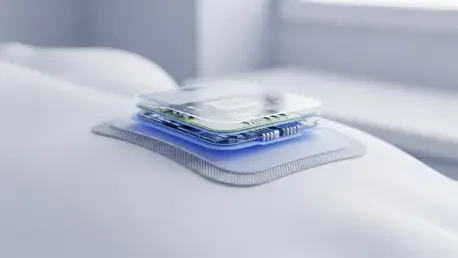
The technological core of this breakthrough is a palm-sized, flexible adhesive patch designed to adhere comfortably to the mother’s abdomen for extended periods. Unlike the rigid, heavy transducers used in traditional hospital settings, this wearable device is engineered using soft, biocompatible materials that move in harmony with the skin. This flexible design is essential for maintaining consistent contact with the maternal body, ensuring that the ultrasound waves can penetrate the tissue without interference from air gaps or shifting sensors. The patch contains a miniaturized array of transducers that are capable of sending and receiving acoustic signals with the same precision as their much larger counterparts. This engineering feat allows for long-term monitoring without the need for a patient to remain stationary or be tethered to a large, complex machine. The result is a device that is as unobtrusive as a standard bandage while providing the diagnostic power of a sophisticated medical imaging system.
To overcome the challenge of constant fetal and maternal movement, which has historically plagued portable ultrasound efforts, researchers developed a sophisticated AI-driven image segmentation algorithm. This software is programmed to identify and lock onto the placental insertion site of the umbilical cord, which serves as a relatively stable anatomical landmark. By focusing on this specific anchor point, the algorithm can maintain a consistent data stream even if the fetus shifts positions or the mother undergoes physiological changes like breathing or walking. The AI was trained on thousands of ultrasound images to recognize the unique patterns of fetal anatomy, allowing it to filter out noise and focus exclusively on the relevant circulatory data. This algorithmic precision ensures that the measurements remain accurate over several hours of monitoring. This automated approach also reduces the need for a technician to manually adjust the probe, making the technology far more accessible.
Clinical Success: Validation and Future Wireless Systems
Clinical validation of the wearable patch involved a study of 62 pregnant participants to ensure that the device met the rigorous safety standards required for obstetric tools. The results demonstrated that the patch’s performance was statistically equivalent to high-end hospital Doppler machines, accurately measuring head circumference and femur length. Furthermore, the acoustic and mechanical energy emissions remained well within the limits established by the American Institute of Ultrasound in Medicine. This safety profile was critical for gaining the trust of both medical providers and patients, as it proved that long-term monitoring did not pose a risk to the developing fetus. The study also highlighted the comfort of the flexible adhesive, which participants were able to wear for several hours without irritation. By proving that hospital-grade diagnostic data could be collected in a wearable format, the research team paved the way for the technology to transition from the laboratory into the delivery room.
The life-saving potential of the ultrasound patch was proven when it detected subtle blood flow fluctuations in a 28-week pregnancy that traditional heart rate monitors had initially missed. By identifying underlying placental dysfunction early, the device allowed the medical team to perform a timely cesarean delivery that saved the infant’s life. Following this clinical success, researchers moved forward with the development of wireless versions to support full-scale outpatient monitoring for high-risk mothers. This evolution enabled patients to remain in their homes while receiving a continuous stream of health data, which significantly reduced hospital admissions and lowered maternal stress. The technology was subsequently applied to other complications such as hypertension and congenital heart disease, expanding its impact across the field of neonatology. These steps ensured that the diagnostic gap in prenatal care was finally closed, providing a sustainable model for future remote healthcare solutions.